|
|
|
The structure of human isocitrate dehydrogenase has not been determined,
but it is known to be composed of three subunits and is allosterically
regulated. The closest homologue has been found in Escherichia coli NADP-dependent
isocitrate dehydrogenase. This homologue only has two subunits and a 13%
identity and 29% similarity based on amino acid sequences. This is not
a close enough homologue to compare the human isocitrate dehydrogenase
to (6). The presence of a distinct gene for IDPm has been observed by the
biochemical characterization, chromosomal location, and molecular cloning
and sequence analyses of IDPm genes in different species (7). Within the
enzyme, an integrated Mg2+ or Ca2+ ion is needed for proper function(6).
Isocitrate dehydrogenase is a functional dimer of 416-residue subunits (9). In yeast, isocitrate dehydrogenase was found to have four binding sites for isocitrate and two each for NAD+, AMP, and Mg2+ or Mn2+ (8). An 11-residue sequence of a subunit with a molecular weight of 39,000 was determined to contain, Ala-Thr-Val-Lys-Gln-Pro-Ser-Ile-Gly-Gly-Tyr; a 16-residue sequence subunit with a 40,000-molecular-weight was, Ala-Thr-Ala-Ala-Gln-Ala-Glu-Gly-Thr-Leu-Pro-Lys-Lys-Tyr-Gly-Gly. There was no heterogeneity at the amino terminus of either subunit, this supported that the subunits were distinct polypeptides. The subunits appear to be immunochemically distinct according to Keys and Henns study on yeast. The enzyme was suggested to have a quaternary structure of a4B4. Another study by Ramachandran and Colman reported a NAD+-specific isocitrate dehydrogenase extracted from a pig heart to have a quaternary structure of a4B2y2 (8). |
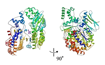
Fig. 1 A closer view of isocitrate dehydrogenase from. Escherichia coli bacteria (16).
|
|
|